
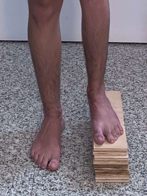
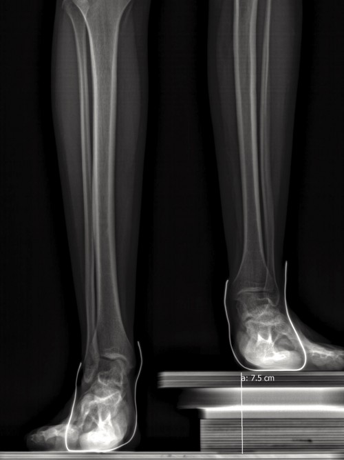
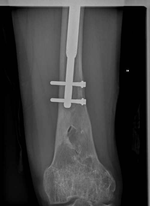
Bone shortenings can reside on one side, creating a difference in length between the two sides. At the level of the upper limbs (arm, forearm) a moderate difference will not have functional consequences. At the level of the lower limbs (thighs, legs), a difference in length, even reduced (2-3 cm) will result in a lameness. In addition, this difference in the length of the lower limbs will cause the pelvis to oscillate from the shorter side, resulting in a scoliotic curvature of the vertebrae with possible lumbar pain. There are different types of shortening of limbs: those that affect only one bone (isolated, intercalary or terminal) and those that affect several adjacent bones (longitudinal shortening). • Congenital short femurs (Proximal Femoral Focal Deficiency) Common, medium or weak length differences are the differences present in the normal population. One person in 1000 has a length difference of 6 cm or more, but several thousand people have a difference between 2 and 6 cm. They may be of congenital origin, in this case increasing gradually from birth to adulthood. The treatment is obviously preferable with endomedullary nails, currently the best hypothesis remains the Guichet nail because it authorizes immediate complete support and sports from 3 to 4 months for elongations of 3 to 4 cm.
In congenital shortenings, there are mainly longitudinal shortenings or shortenings of a predominant bone (e.g. femur), in which the other bones are minimally affected (e.g. tibia and foot). In traumatology, shortenings are generally isolated within a single bone, resulting either from initial shortening or growth changes induced by initial trauma.
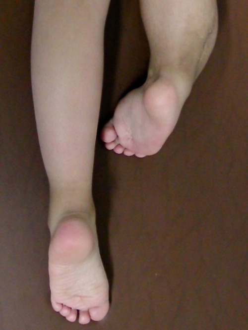
Among the congenital unilateral shortenings of the lower limbs in relation to the affected bone, there are :
• Femimelie peroniarie
• Tibial hemimelias
• Foot abnormalities causing deformation (e.g. varo-equine foot or twisted foot)
TYPES OF INJURIES TREATMENT JOINT CORRECTIONS LENGTHENING
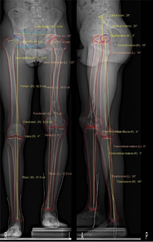
It is an anomaly included in the longitudinal deficits of the lower limbs. This means that a limb is often deficient on the outside (deficit of the femur, anterior-lateral cruciate ligament, and peroneal hemimelia). Depending on the main problem (femur or leg), the symptomatology will be mainly found on this segment, but it may also be minimally found on other segments. In this way, a short congenital femur can associate an important shortening of the femur, a deficit of the anterior-lateral cruciate ligament (absence), a deformation of the lateral condyle and a shortening of the leg from moderate to weak.
The objective of the treatment, after a precise and complete evaluation and the study of the possibilities of reconstruction, is to obtain functional joints and a normal limb length at the end of the elongation. This is possible if the patient has not undergone an excessive number of surgical operations previously.
First of all we need to correct and stabilize the joints. The elongations are carried out later, limiting the number of surgical procedures to the minimum necessary.
An axis and good joint stability are indispensable before any elongation. Joint corrections for axle defect or pseudoarthrosis are made as needed. Often, there is an absence of knee ligaments that can be healed with an operation to stabilize the joint before stretching.
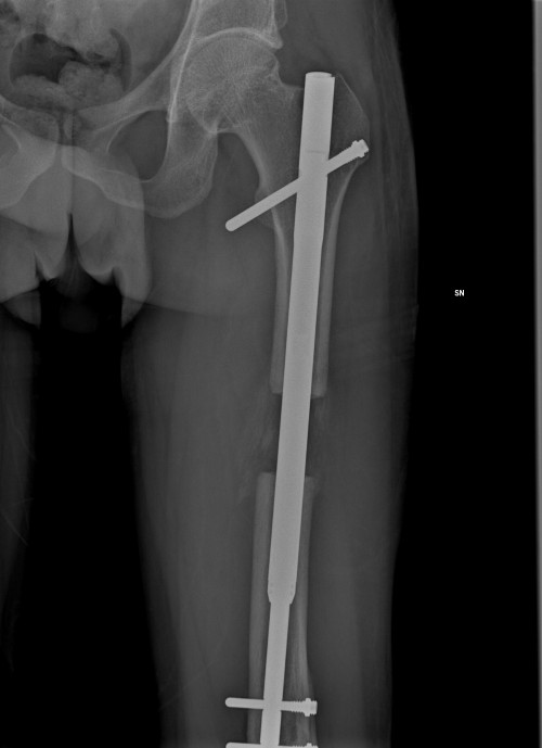
Elongations pose less risk when they are made by means of elongation nails than when they are made by means of external fasteners. Axis corrections can be performed during the same surgery. It is possible to take care of the patient at the end of the growth for shortening from 12 to 18 cm. Meanwhile, orthopaedic shoes or prosthetics can be used.
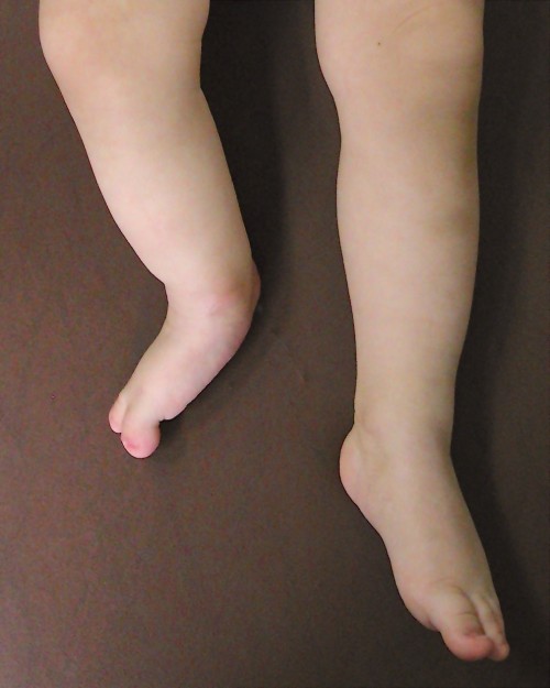
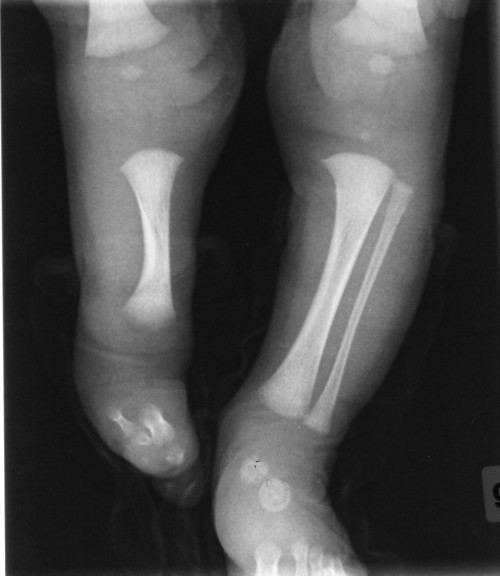
Fibular hemimelia is a congenital deformation that results in the lack of one or more bone segments in the lower limbs. It can be classified in two types: - longitudinal, where there is a missing radius of medial or lateral bone of one limb, or one between ulna and radium, or between tibia and fibula. - transverse, when the distal part of a limb is completely missing and a stump may be present reminiscent of that of an amputation. For example in the case of the foot when there is an absence or partial deficit of the fifth finger.
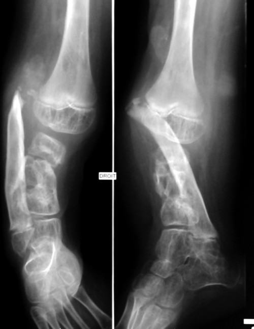
The tibial hemimelia is a rare congenital anomaly with a tibia deficiency coupled with a relatively intact fibula. This pathology can be unilateral or bilateral, it can present an isolated defect or more musculoskeletal malformations. The goal is to restore the functionality of the limb so as to allow walking, performing sports activities, driving a car, etc. The treatment is very complex and the planning must be carefully carried out to avoid any therapeutic error. A fusion of the knee with a considerable elongation, especially of the femur, will determine a supporting limb, long but not functional. The function has been privileged to the complete correction of the stature, causing the loss of function. It is necessary to be assisted very soon in a competent centre.
Weymouth Street Hospital
42-46 Weymouth St.
London W1G 6DR, UK
Phoenix Hospital Group
9 Harley Street
London W1G 9QY, UK
Harley Street Specialist Hospital
18-22 Queen Anne St.
London W1G 8HU, UK
Studio Dr. Guichet
Corso Magenta, 44
20123 Milano, IT
+39 0236758514